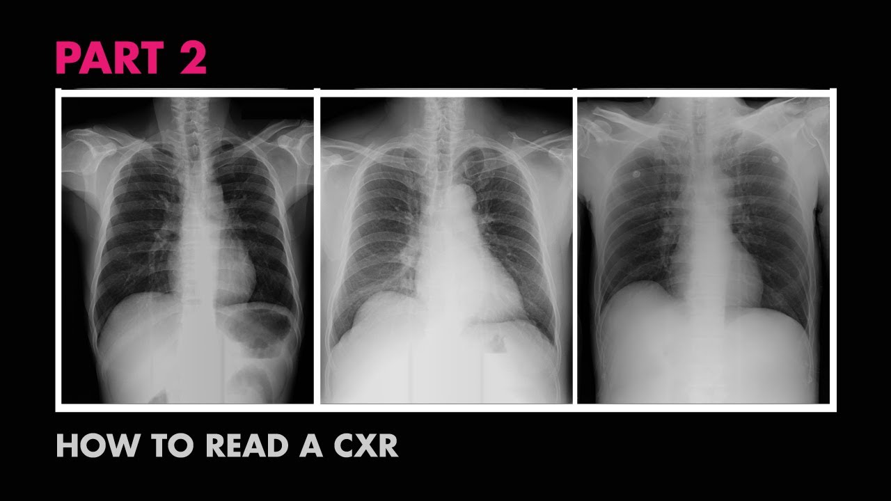
For every thousand photons reaching the patient 100200 photons are scattered. Nine pairs of ribs should be seen posteriorly in order to consider a chest x-ray adequate in terms of inspiration.
 How To Read A Chest X Ray With Pictures Wikihow
Much can be said about specific diagnoses that can be made on plain imaging but this post is more about an approach to a general MSK x-ray whether it is a study of the fingers hands wrists forearms humeri shoulders.
How to read x ray. This course will help you start from the very beginning till you are able to read any CXR. In this example the x-ray tube is emitting photons which are either reflected or absorbed by the patient. For chest X-Rays there is a classic schematic.
In this introductory video we talk about how dental x-rays work how to read them and how to apply the buccal object rule to localize objects in an x-ray i. This is the radiation dose. If the crystal size is too small it can determine sample composition crystallinity and phase purity.
The rest are absorbed by the patient. This technique sends x-ray beams through it. But the basics of Chest Xray here will guide you through various aspects including Counting ribs PA vs AP view Inspiratory vs Expiratory Xray Erect vs Supine Lucency and Opacity and some common.
X ray is a type of radiography and most widely used investigation. It first appears too complicated to read the chest xrays because we barely know what lies where and what to make out of it. The patient will place their chest against a plate which digitally records the image.
Measure the distance from the medial end of each clavicle to the spinous process of the vertebra at the same level which should be equal. Chest X-Ray is the most widely practiced imaging procedure and knowing how to read it is a basic skill for every medical student or practitioner. ABCDEF You should first check the patients name and date of the film.
How To Read A Plain Film Chest X-Ray There is no right or wrong way to read a chest x-ray but it is beneficial to develop a systematic method of doing so. Patient Data name history age sex old films Routine Technique. X-ray diffraction is a common technique that determine a samples composition or crystalline structure.
With the lateral view the patient stands with the left side pressed again the plate. Name and date of birth of the patient Side of extremitybody Date of x-ray Two views help to fully describe the fracture in both planes. The red arrows are pointing to the pulp where the nerve and blood vessels are located for a tooth.
The first in our c. Abnormal shadowing or lucency. Information found on the x-ray are.
Just 20 reach the image detector. Midline or deviated caliber mass. APPA exposure rotation supine or erect.
The blue arrows are pointing to the healthy enamel. X-rays of the two adjacent joints must be taken. How to Read a Chest X-Ray.
Reading plain x-ray images of the skeleton is a basic radiology skill. The first thing to always check is that the film is associated with the correct patient. We will start with how to assess the four basic pillars of image quality using the mnemonic RIPE.
The purple arrows are pointing to areas of decay which show up as a dark spot on x-rays. Turn off stray lights optimize room lighting view images in order. After doing so to read a PA view I utilize a mnemonic called RIP ABCDEFGH.
Just a fraction of the x-rays pass through the patient to the image intensifier. Finally you should check patients position such as supine erect or semi-erect. The X-ray tube is position six feet away from the patient.
Other less common views might include an AP view which can be done in a hospital with a mobile X-ray unit. A close second to reading chest x-rays learning how to read a musculoskeletal image is an essential skill to have. For larger crystals such as macromolecules and inorganic compounds it can be used to determine the structure of atoms within the sample.
It is easy to miss a fracture with only one view see red circle. You should also check the side marker and the film position PA or AP. The dentin layer is between the enamel and the pulp.
Just a shaft view is not enough. First name surname date of birth.


